Accelerate AI-powered pathology research with access to high-quality whole slide images (WSI) linked to rich clinical, molecular, and outcomes data. Discovery’s WSI library enables the development of machine learning models for cancer detection, biomarker discovery, patient stratification, and treatment-response prediction.
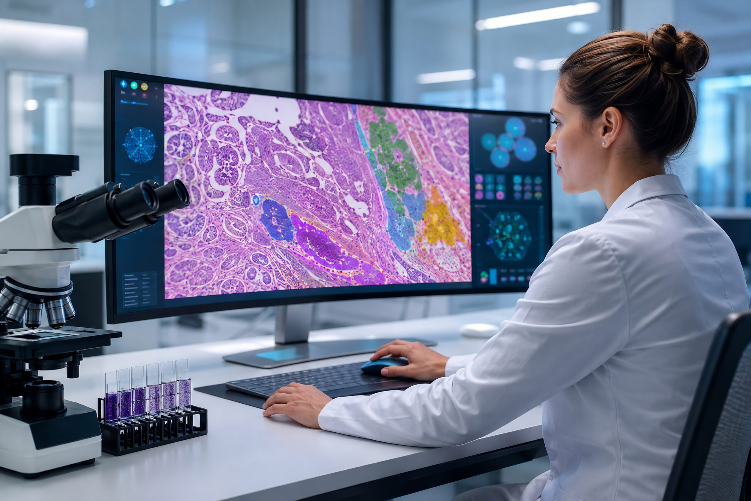
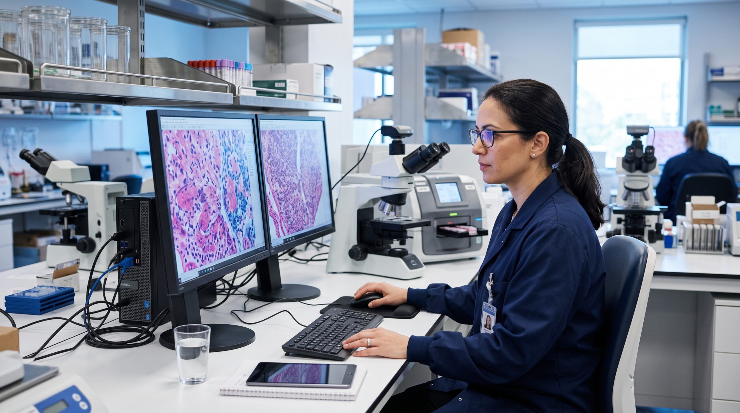
Discovery’s WSI library is designed for AI and digital pathology applications, each dataset combines high-resolution tissue images with curated metadata to support robust model development and validation.
ArtifactDetect and TumorDetect are research use only products available on PathAI’s AISight® Image Management System, not for use in diagnostic procedures.
| Image Only | Image + FFPE | Total Images | |
|---|---|---|---|
|
Lung Cancer |
43,000 |
7,000 |
50,000 |
|
SpecimenSeqTM FFPE Tissues Targeted NGS Solid Tumor Panel Results |
33,500 |
7,500 |
41,000 |
|
Patient Follow-Up Data Treatment and Outcome Summaries |
1,300 |
300 |
1,600 |
|
Additional Characterizations MSI, MMR, PD-L1, HPV, HLA, and More |
20,000 |
6,000 |
26,000 |
Discovery’s WSI library is continuously updated with new images; therefore, the numbers shown may change. This table reflects only a subset of Discovery’s total biospecimen inventory.
Enhance image-based analysis with molecular and longitudinal clinical data.
Targeted NGS panel results available from:
Available cohorts may include:
Selected cohorts include up to 2 years of follow-up data, with patient treatment and outcome summaries at:
Extend the value of your tissue images with industry-leading digital pathology and AI-powered analysis services.
Leverage PathAI technology to rapidly assess and annotate whole slide images at scale.
Transform your physical tissue slide archives into a searchable, scalable digital asset library.
Unlock deeper biological insights with the full suite of PathAI analytical capabilities.
Access expert pathology support for annotation, validation, and quality assurance needs.
Successful AI models depend on access to high-quality, well-annotated, and clinically relevant data. AI-ready datasets typically combine high-resolution whole slide images with standardized quality control, verified annotations, comprehensive metadata, and linked clinical, molecular, and outcomes information.
Discovery's WSI datasets are curated to support a broad range of digital pathology and precision medicine applications, including:
By combining high-quality whole slide images with rich contextual data, our customers can develop more accurate models and uncover insights that would be difficult to identify from image data alone.
While image data alone can provide valuable insights, combining pathology images with molecular, biomarker, clinical, and outcomes data allows researchers to build more powerful models. Multi-modal approaches can uncover relationships between tissue morphology, genomic alterations, treatment response, and patient outcomes that may not be apparent from any single data type.
Slide bank digitization converts physical tissue archives into research-ready digital assets that are easier to store, access, analyze, and share. Organizations often digitize slides to preserve valuable specimens, support remote pathology workflows, enable AI-driven analysis, and maximize the long-term value of their tissue collections.
Many AI models are trained to recognize what is present in a tissue sample today. Outcomes data adds information about what happened to the patient over time, allowing developers to create models that predict treatment response, disease recurrence, progression, and survival. This can significantly increase the clinical utility of AI algorithms.
