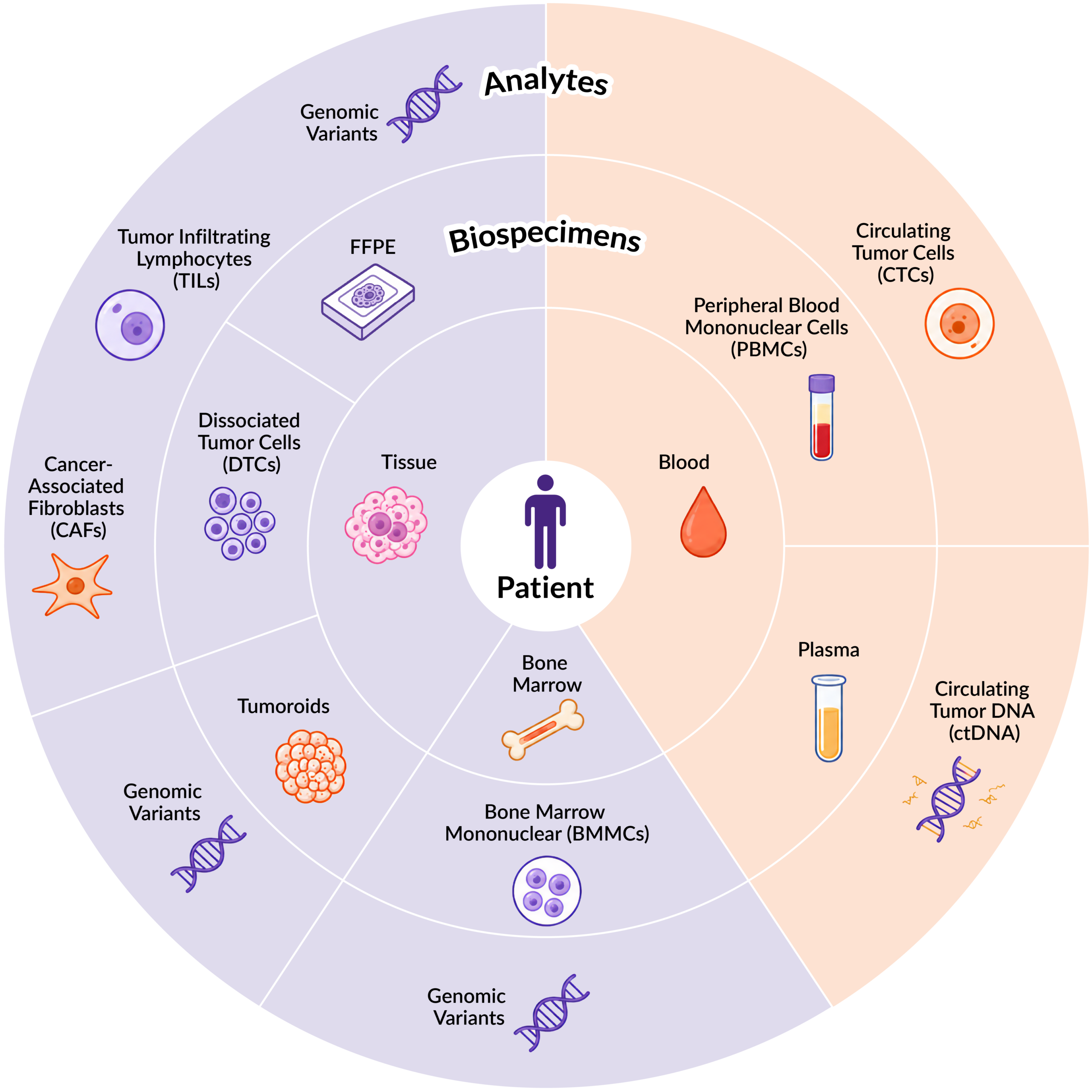
In the final post of our analyte blog series, we take a look at circulating tumor cells (CTCs, Figure 1). CTCs are intact cancer cells that break away from a primary tumor, enter the bloodstream, travel to other sites and have the potential to establish secondary lesions.
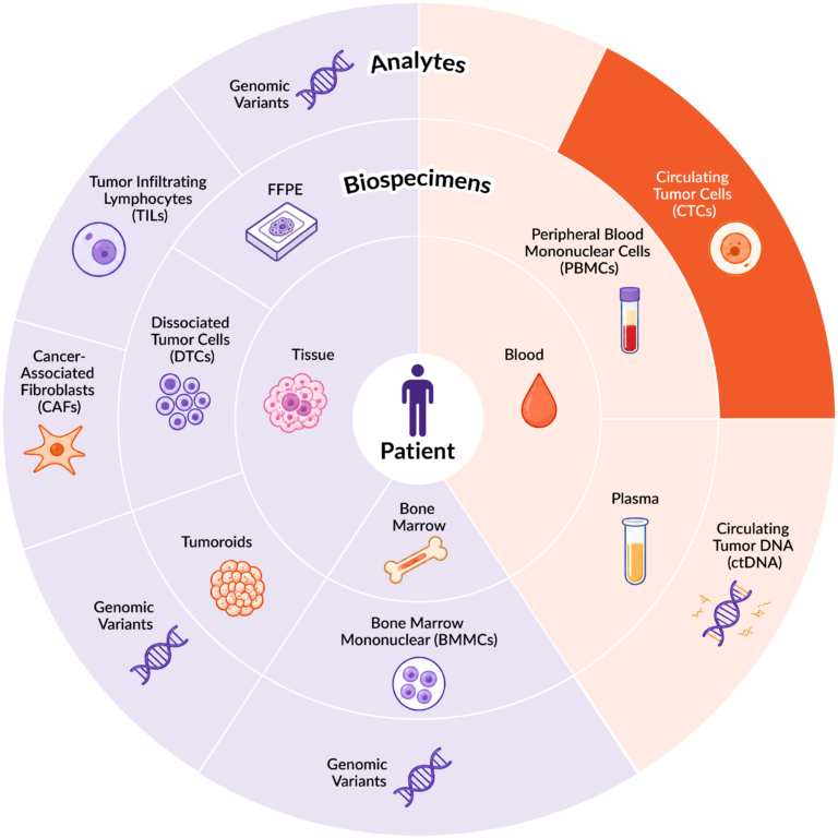
Figure 1. Distribution of key analytes across different biospecimen sample types.
Because CTCs are closely linked to the metastatic process, their presence and abundance in blood can provide valuable prognostic information about disease progression and patient outcomes. For example, the number of CTCs can be used to differentiate between cancers at various stages. Generally speaking, patients with late-stage or more advanced disease tend to have a higher abundance of CTCs than patients with earlier-stage disease1. However, CTCs are extremely rare, which presents an analytical challenge. It is estimated that only one CTC is present among approximately 106to 108 leukocytes2. Therefore, the ability to isolate them from millions of blood cells is essential for unlocking their value as a window into metastatic disease.
A Needle in a Haystack: Isolating CTCs from Peripheral Blood
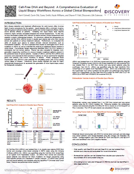
A variety of approaches have been developed to enrich and isolate CTCs, including physical separation based on cell size, density, deformability, or the electrical properties of the cell membrane. Immunomagnetic antibody-based capture methods can also be used to enrich CTCs based on the expression of cell-surface markers, such as epithelial cell adhesion molecule (EpCAM). Figure 2 illustrates this approach, showing the identification of EpCAM+ CTCs within the CD45– fraction of peripheral blood mononuclear cells (PBMCs) samples obtained from a normal donor and patients with early- or late-stage lung cancer. As expected, very few CTCs are detectable, even in the late-stage lung cancer patient.
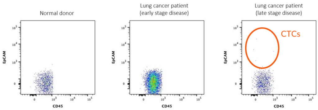
Figure 2. PBMCs from a normal donor and patients with early- and late-stage lung cancer were lineage-depleted using a hematopoietic lineage depletion kit. Lineage-negative cells were stained for CD45 and EpCAM, and EpCAM+ CTCs were identified in the CD45– fraction.
As interest in CTC analysis continues to grow, researchers are developing increasingly sophisticated approaches, including microfluidic and size-based platforms, to improve the recovery of these rare cells while preserving their viability and biological characteristics2,3.
While peripheral blood is the most common sample type for CTC analysis, CTCs can also be isolated from other biofluids. In patients with metastatic lung adenocarcinoma (ACA), CTCs may be present in pleural effusions, while in patients with ovarian cancer, CTCs may also be recovered from ascites fluid within the peritoneal cavity4,5.
Studying tumor cells from sites beyond the primary tumor can provide important insights into the biology of metastasis. CTCs often have a different genetic makeup from the primary tumor and may more accurately reflect how cancer cells mutate and evolve as the disease progresses and spreads to new sites6. In this context, access to matched samples from the same patient enables researchers to compare tumor cells across disease compartments. For example, CTCs isolated from PBMCs and dissociated tumor and tissue cells (DTCs) from the same patient can be compared to identify genetic drivers that contribute to metastasis, which could help researchers identify novel biomarkers for early disease detection or help develop new therapeutic strategies.
Liquid Biopsy
As we described in blog 2, liquid biopsy is a minimally invasive sampling technique, which allows for the analysis of circulating tumor-derived material, including CTCs, cell-free circulating tumor DNA (ctDNA) and extracellular vesicles (EVs), in body fluids, primary peripheral blood7. Because blood can be collected at multiple timepoints, liquid biopsy also enables longitudinal monitoring of tumor-derived biomarkers throughout disease progression and treatment.
The number of CTCs can provide valuable insights into tumor progression and therapeutic response. In many solid tumors, including metastatic breast, prostate, and lung cancers, elevated baseline CTC numbers are strongly associated with shorter progression-free and overall survival1,2. A decrease in CTC numbers during or after treatment, including chemotherapy, targeted therapy, or surgery, can indicate a favorable treatment response. In contrast, an increase or persistence of high CTC levels during treatment can be associated with treatment failure, disease progression, or a higher risk of recurrence, sometimes appearing earlier than changes detected by standard imaging or traditional blood-based tumor markers.
Additionally, CTC analysis can provide information beyond cell enumeration. In contrast to ctDNA and EVs, CTCs are intact, viable cells that can support functional characterization7. Genomic, transcriptomic, proteomic, and epigenetic profiling of CTCs provides unique opportunities to investigate tumor biology and the metastatic cascade at the molecular level7-8. This multidimensional characterization has significant implications for personalized medicine, enabling researchers to track mutation status, identify biomarkers, and investigate how tumor characteristics change over time and relate to disease progression and treatment response.
From Analyte to Insight
Across this series of articles, we have explored how different tumor-derived analytes can help answer different questions about tumor biology, disease progression, and treatment response. Each analyte brings unique characteristics and analytical opportunities to advance diagnostic and therapeutic development.
At Discovery Life Sciences, we know that high-quality, clinically characterized biospecimens are the foundation for meaningful analyte research. That’s why we have built a deep and diverse biobank of clinically annotated biospecimens that span a broad range of cancer types and disease stages, enabling researchers to investigate cancer from multiple biological perspectives and uncover insights that move cancer research forward.
References
- Nguyen, T. N. A., Huang, P. S., Chu, P. Y., Hsieh, C. H., & Wu, M. H. (2023). Recent Progress in Enhanced Cancer Diagnosis, Prognosis, and Monitoring Using a Combined Analysis of the Number of Circulating Tumor Cells (CTCs) and Other Clinical Parameters. Cancers, 15(22), 5372.https://doi.org/10.3390/cancers15225372
- Dai, C. S., Mishra, A., Edd, J., Toner, M., Maheswaran, S., & Haber, D. A. (2025). Circulating tumor cells: Blood-based detection, molecular biology, and clinical applications.Cancer Cell, 43(8), 1399–1422. https://doi.org/10.1016/j.ccell.2025.07.008
- 3.Vidlarova, M., Rehulkova, A., Stejskal, P.,Prokopova, A., Slavik, H., Hajduch, M., & Srovnal, J. (2023). Recent Advances in Methods for Circulating Tumor Cell Detection. International journal of molecular sciences, 24(4), 3902. https://doi.org/10.3390/ijms24043902
- 4.Zhu, Y., Allard, G. M., Ericson, N. G., George, T. C., Kunder, C. A., & Lowe, A. C. (2021). Identification and characterization of effusion tumor cells (ETCs) from remnant pleural effusion specimens. Cancer Cytopathology, 129(11), 893–906.https://doi.org/10.1002/cncy.22483
- 5.Nunes, D., & Ricardo, S. (2022). Ovarian Cancer Ascites as a Liquid Tumor Microenvironment. Ovarian Cancer, 43–55.https://doi.org/10.36255/exon-publications-ovarian-cancer-tumor-microenvironment
- 6.Kim, W., Cho, S., Lee, J., Lee, J., Ji, S., Sung, H., Jung, W., Jeon, J. H., Kim, K., & Jheon, S. (2024). Mutational differences between primary cancer tissue and circulating tumor cells in early-stage non-small cell lung cancer. Translational lung cancer research, 13(11), 3026–3038.https://doi.org/10.21037/tlcr-24-709
- 7.Smit, D. J., & Pantel, K. (2024). Circulating tumor cells as liquid biopsy markers in cancer patients.Molecular Aspects of Medicine, 96, 101258. https://doi.org/10.1016/j.mam.2024.101258
- 8.Ma, L., Guo, H., Zhao, Y., Liu, Z., Wang, C., Bu, J., Sun, T., & Wei, J. (2024).Liquid biopsy in cancer current: status, challenges and future prospects. Signal transduction and targeted therapy, 9(1), 336. https://doi.org/10.1038/s41392-024-02021-w

Debbie King
Scientific Writer, Discovery Life Sciences