In the first post of this series, the analyte of interest was genomic variants, which can be detected across multiple biospecimen types. As we discussed, the optimal sample for detection depends on the biological question being asked, reflecting the complexity of their distribution across different biological matrices. For other analytes, however, where they live is more straightforward.
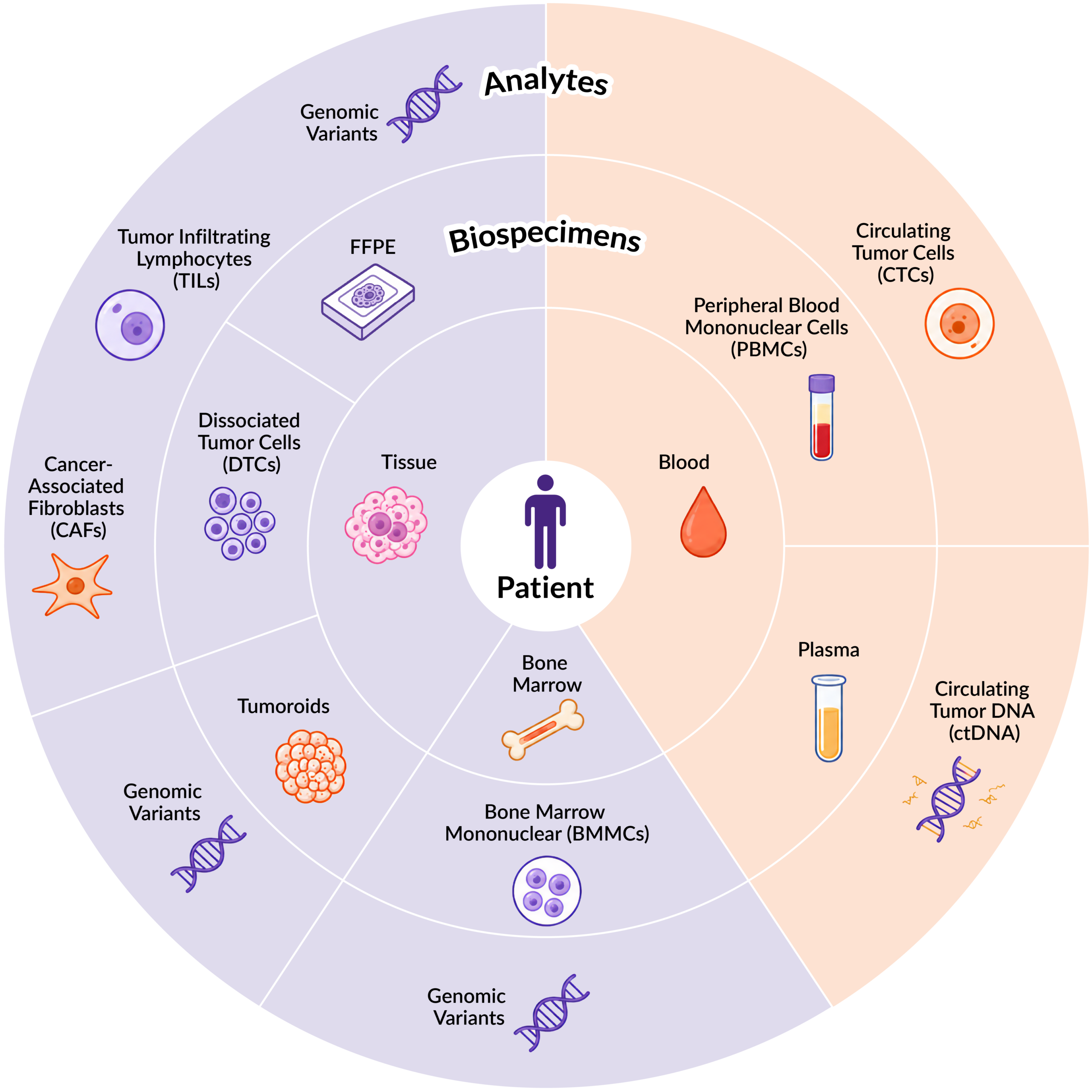
In this post, we focus on cell-free DNA (cfDNA), an analyte primarily found in biofluids. cfDNA are short and long fragments of DNA that are released into circulation as cells undergo apoptosis, necrosis, and other cellular processes (Figure 1). These fragments carry the genetic information of the cells they came from, which means cfDNA analysis can provide a minimally invasive view into biological processes throughout the body.
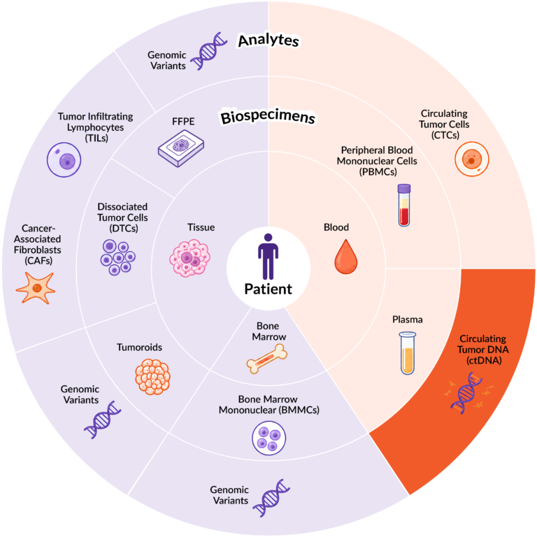
Figure 1. Distribution of key analytes across different biospecimen sample types.
In cancer, a portion of circulating cfDNA comes from tumor cells. This tumor-derived fraction is called circulating tumor DNA (ctDNA). Because ctDNA contains cancer-related genetic and epigenetic changes of the tumor cells, it can be used to study tumor biology, support treatment decisions, monitor treatment response and resistance, track minimal residual disease, and help with early cancer detection1.
cfDNA-based liquid biopsy is becoming more widely used because it is generally well tolerated and far less invasive than traditional tissue sampling. Therefore, understanding where cfDNA originates and the variables that impact its detection is essential for generating accurate and biologically meaningful insights.
Where Is Cell-Free DNA Found?
cfDNA can be detected in a variety of biofluids, including blood, urine, cerebrospinal fluid (CSF), saliva, and pleural effusions. Among these, blood remains the most widely used and well-characterized source for cfDNA analysis, because it is accessible and routinely collected as part of clinical patient care. Blood is also highly biologically rich, reflecting molecular contributions from tissues and organs throughout the body, whereas other biofluids often provide more localized biological signals.
cfDNA is a low-abundance analyte, with concentrations typically ranging from 1 to 10 ng/mL in healthy individuals2. At those levels, pre-analytical variables can have a major effect on sample quality and downstream analysis. Blood collection tube type, processing time, and plasma preparation methods can all influence the amount and integrity of cfDNA that can be recovered.
Blood Collection Tube Type
Whole blood is typically collected in ethylenediaminetetraacetic acid (EDTA) or Cell-Free DNA BCT® (Streck) tubes, which contain additives that help preserve blood cell integrity after collection to minimize the release of contaminating genomic DNA into plasma. Following collection, blood is centrifuged to generate double-spun plasma (DSP), the cell-free fraction used for cfDNA analysis.
The choice of collection tube determines how the blood sample remains suitable for processing to isolate cfDNA. Streck tubes contain a preservative that stabilizes nucleated blood cells and can maintain sample stability for more than 48 hours before processing. EDTA tubes, by comparison, are less stable and need to be processed as quickly as possible to limit genomic DNA contamination from blood cells. In Discovery’s workflow, EDTA-collected samples are processed within two hours of collection and Streck-collected samples within 48 hours to maximize cfDNA recovery while minimizing genomic DNA contamination.
In our evaluation, when EDTA-collected samples are processed promptly, comparable levels of cfDNA can be isolated from DSP prepared using either tube type.
For cfDNA studies with specific program requirements, we can prospectively collect blood using custom collection tube types to meet downstream assay requirements.
Single vs. Double-Spun Plasma
Single-spun plasma contains cfDNA, but it can also carry substantial amounts of contaminating cellular and genomic DNA that may interfere with downstream analysis. For this reason, double-spun plasma is generally preferred for cfDNA work as the second centrifugation step helps remove residual blood cells and cellular debris, improving the ability to detect low-abundance cfDNA.
However, many existing plasma collections, particularly those generated for studies not originally designed for cfDNA analysis, may only be available as single-spun plasma. We have found that single-spun plasma samples collected from solid tumor patients can be processed again to generate DSP that is more suitable for cfDNA analysis. Subjecting single-spun plasma to an additional centrifugation step to produce DSP effectively removes the contaminating material resulting in a higher concentration of detectable cfDNA, as seen in Figure 2, thereby expanding the utility of existing plasma collections for downstream cfDNA applications.
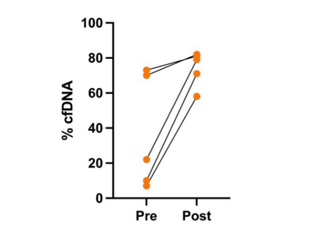
Figure 2. % cfDNA levels in single spun plasma samples from 5 donors before (pre-) and after (post-) an additional centrifugation step to generate DSP.
Detection in Other Biofluids
Although plasma remains the gold standard for cfDNA analysis, other biofluids are increasingly being explored for specific applications. Among these, urine has emerged as a promising non-invasive source of cfDNA that enables repeated sampling with minimal burden to patients. Additionally, the lower protein concentration in urine resulting from kidney filtration simplifies cfDNA isolation, which reduces the analytical complexity compared to cfDNA from blood samples3.
In urological cancers in particular, urine cfDNA is of growing interest as a disease biomarker. Beyond urological applications, urine-based cfDNA analysis is also being investigated for broader oncology and non-oncology use cases to drive further progress in disease detection and treatment monitoring.
Figure 3 shows that cfDNA can be isolated from both normal and disease-state urine samples, with higher average levels observed in the disease cohort, consistent with increased cellular turnover and tumor-derived DNA shedding.
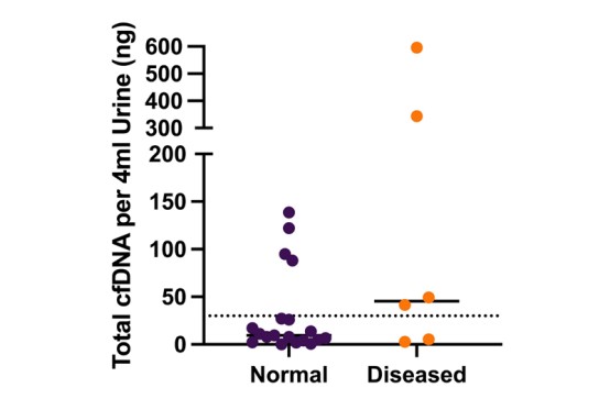
Figure 3. Quantification of total cfDNA (ng) isolated from urine samples collected in Streck tubes from 19 normal and 6 diseased patients. The average total cfDNA of the normal cohort was 30.8 ng compared to 173.2 ng in the diseased cohort.
cfDNA and Liquid Biopsies
Liquid biopsy tests are designed to detect low levels of circulating cfDNA for diagnostic and prognostic purposes. In oncology, these tests often focus on the even smaller fraction of cfDNA released by cancer cells into plasma as ctDNA. Several assays have already received regulatory approval, highlighting the growing clinical value of this approach. One example is the Roche cobas® EGFR Mutation Test v2, which detects specific EGFR mutations in plasma to support targeted therapy decisions in non-small cell lung cancer.
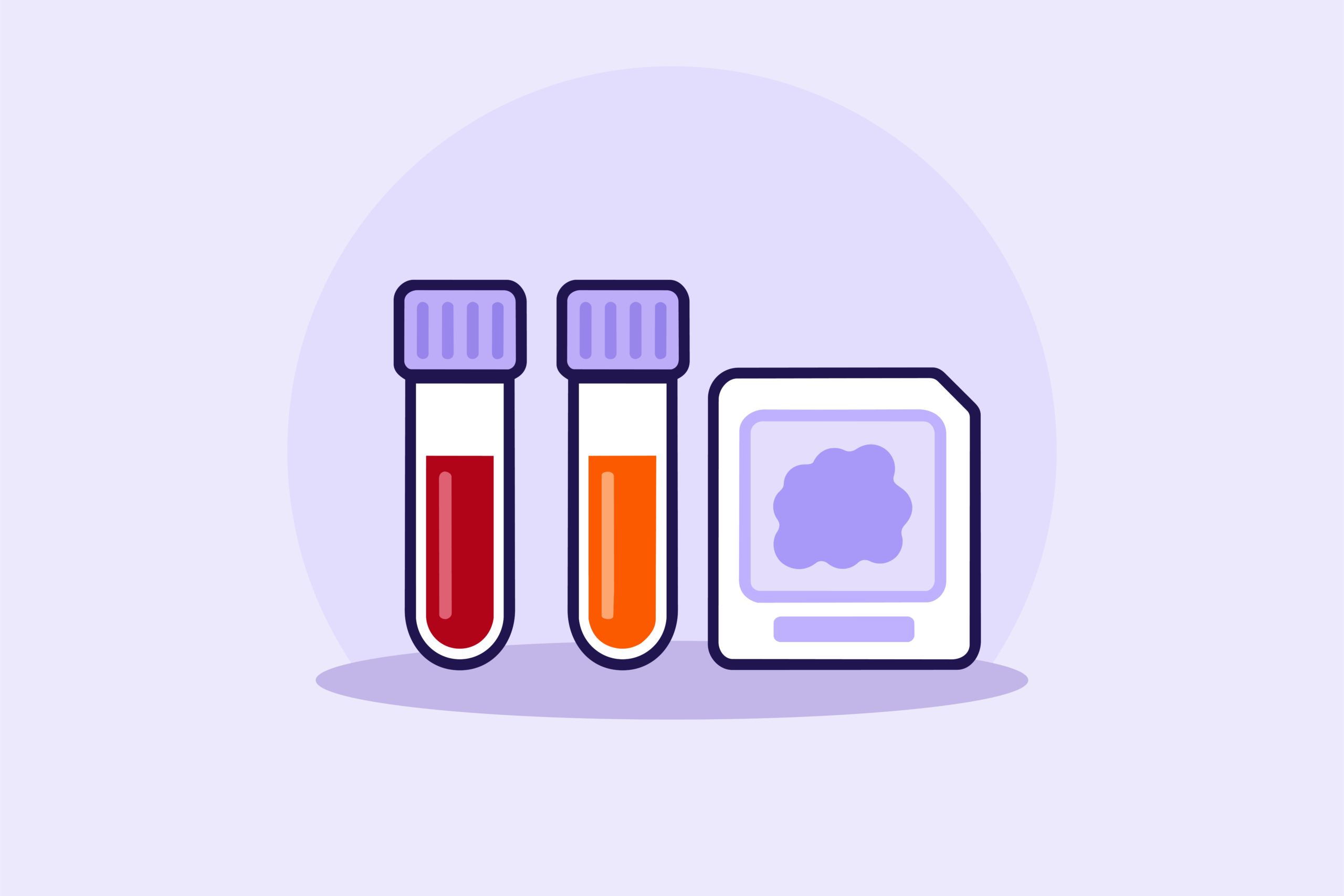
To validate a plasma liquid biopsy assay, cfDNA findings are typically compared with matched tissue samples, most often formalin-fixed, paraffin-embedded (FFPE) tissue, to confirm concordance between circulating and tissue-derived signals. Matched buffy coat analysis is also often included to account for clonal hematopoiesis of indeterminate potential (CHIP), which can introduce somatic variants from hematopoietic cells and complicate interpretation of cfDNA results.
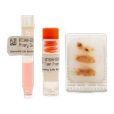
Discovery Life Sciences’ biospecimen portfolio includes a range of matched biospecimen sets to support these initiatives, including FFPE, DSP, and buffy coat samples collected from the same patient. Matched urine, DSP, buffy coat, and FFPE sample sets can also be used for the development and validation of emerging urine-based liquid biopsy assays. And because our biospecimen inventory sits alongside our genomics and molecular pathology labs, we can help maximize the value of matched samples by enabling integrated biospecimen characterization and molecular analysis, including sequencing, methylation profiling, immunohistochemistry (IHC), and other downstream characterization within an integrated workflow.
As liquid biopsy technologies continue to mature, cfDNA is becoming an increasingly important analyte for disease detection, monitoring, treatment selection, and patient stratification across oncology and beyond. But like any analyte, its value depends on selecting the appropriate sample type and handling it correctly from the start.
More to Come in the Series…
Stay tuned for the next blog in this series where we will explore circulating tumor cells (CTCs) as another analyte found within the blood-based biospecimen landscape.
References
- MouliereF. (2022). A hitchhiker’s guide to cell-free DNA biology. Neuro-oncology advances, 4(Suppl 2), ii6–ii14. https://doi.org/10.1093/noajnl/vdac066
- Dao, J., Conway, P. J., Subramani, B., Meyyappan, D., Russell, S., & Mahadevan, D. (2023). Using cfDNA and ctDNA as Oncologic Markers: A Path to Clinical Validation.International journal of molecular sciences, 24(17), 13219. https://doi.org/10.3390/ijms241713219
- Salfer, B., Li, F., Wong, D. T. W., & Zhang, L. (2022). Urinary Cell-Free DNA in Liquid Biopsy and Cancer Management. Clinicalchemistry, 68(12), 1493–1501. https://doi.org/10.1093/clinchem/hvac122

Debbie King
Scientific Writer, Discovery Life Sciences