We are halfway through our blog series, in which we map key analytes to the biospecimens where they can be found. After exploring genomic variants and cell-free DNA in earlier posts, we take an in-depth look at tumor-infiltrating lymphocytes (TILs).
TILs are immune cells that infiltrate and reside within the tumor microenvironment (TME), most commonly including T-cell populations such as CD8+ cytotoxic T cells, CD4+ helper T cells, and regulatory T cells that contribute to anti-tumor immune responses (Figure 1). Because TIL abundance, phenotype, and spatial distribution can reflect the state of the anti-tumor immune response, TILs are widely studied as biomarkers of immunotherapy response1,2.
They are also clinically relevant both as targets for evaluating immunomodulatory therapies, such as immune checkpoint inhibitors, and as therapeutic starting material in adoptive cell therapies, including the FDA-approved TIL therapy lifileucel (brand name Amtagvi) for the treatment of advanced melanoma2.
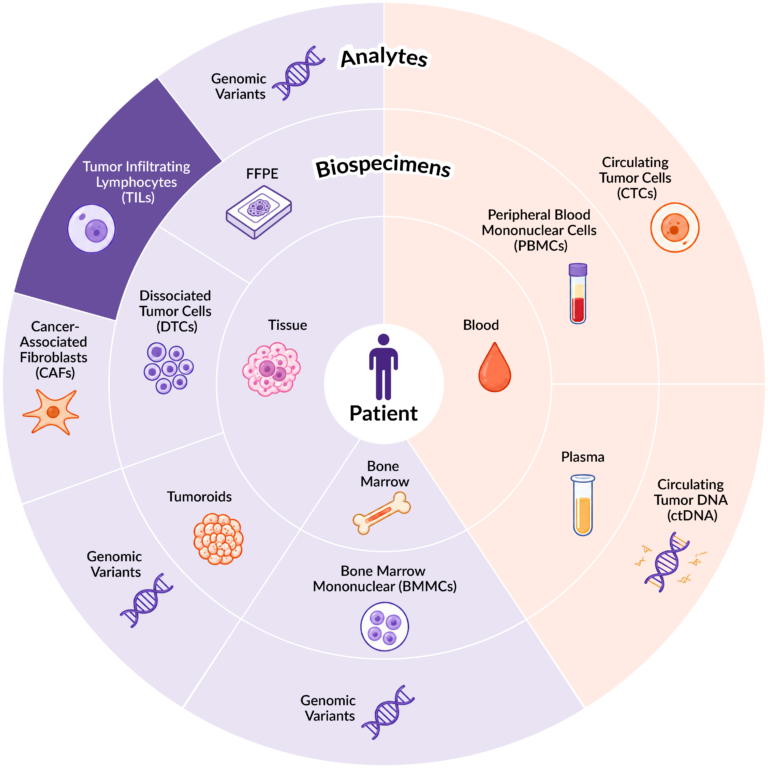
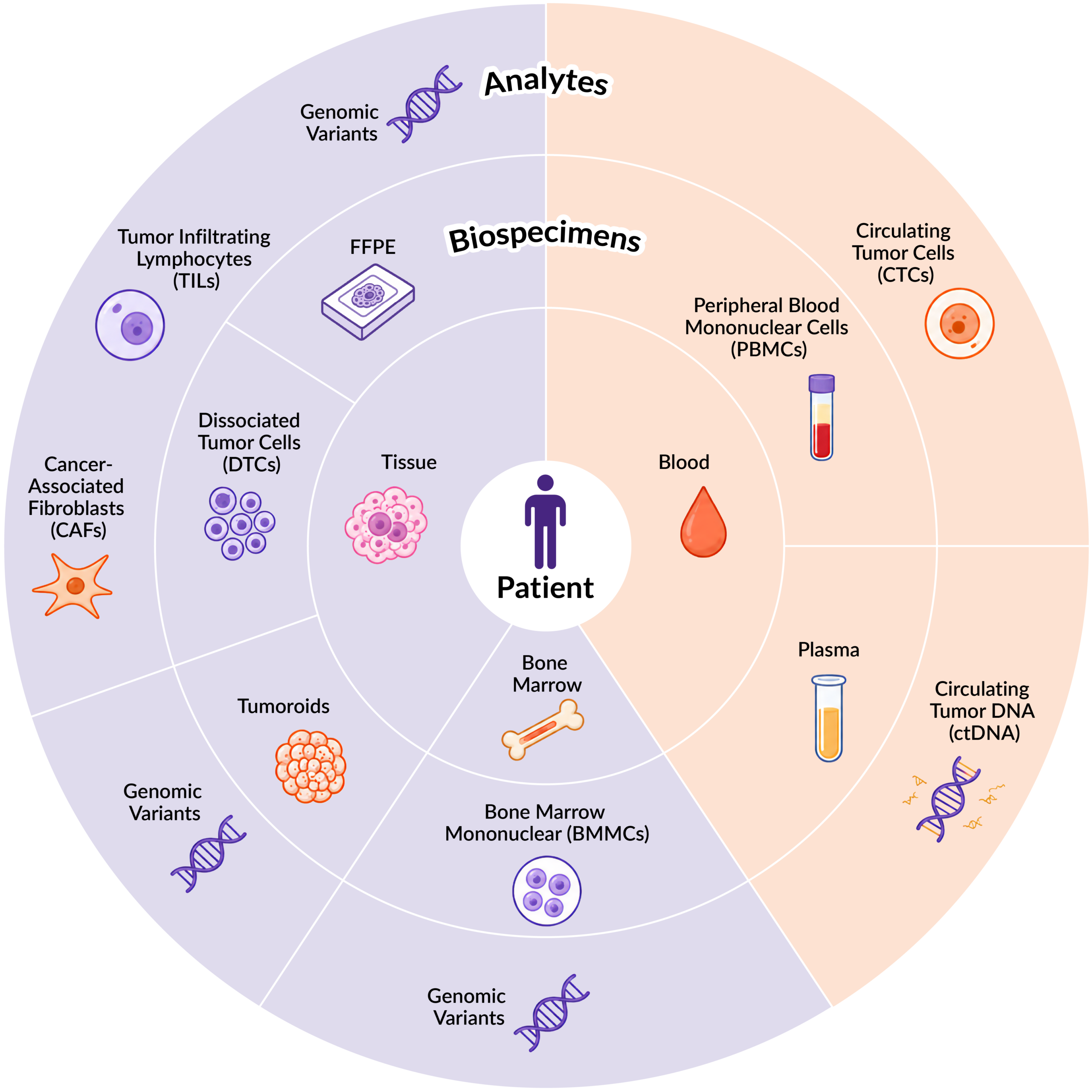
Figure 1. Distribution of key analytes across different biospecimen sample types.
So where should researchers look when TILs are the analyte of interest?
When researchers need viable immune cells for isolation, enrichment, and downstream functional analysis, dissociated tumor and tissue cells (DTCs) are an important biospecimen type to consider. These viable single-cell suspensions are generated from patient tumor tissues through a mechanical and enzymatic dissociation technique. DTCs contain a heterogeneous mix of cell populations within the TME, including tumor cells, stromal cells, and TILs.
What You Need to Know About Dissociated Tumor and Tissue Cells (DTCs)
The complex cellular composition of DTCs creates opportunities to characterize and isolate specific cell subpopulations within the tumor microenvironment for downstream analysis. Flow cytometry can be used to profile the cellular makeup of a DTC sample, while immunomagnetic separation can enrich or purify selected cell populations for downstream applications. The pan-leukocyte surface marker CD45 is especially useful when studying TILs because it is expressed on most immune cells. Quantifying CD45+ cells provides an initial assessment of the immune-cell contribution within a DTC sample.
Notably, the immune cell component of a DTC sample can vary considerably between tumor types and even among patients with the same indication. This variability is also observed across DTC biospecimens within the Discovery Life Sciences (Discovery) biobank. In our analysis, DTCs isolated from different patients across multiple oncology indications demonstrated substantial variability in CD45+ immune cell frequency. The highest percentages of CD45+ cells were observed in cervical, head and neck, kidney, liver, and lung tumor samples, while bladder, brain, and melanoma specimens contained lower proportions of immune cells (Figure 2)3.
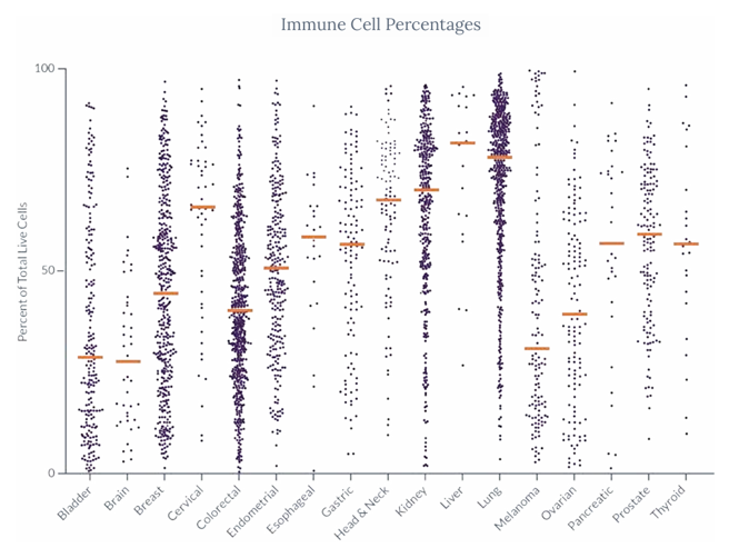
Figure 2. The percentage of CD45+ immune cells within DTCs across seventeen unique oncology indications. Each dot represents a unique patient collection, with the orange bars representing the average tumor percentage per indication.
As part of the QC process, each lot of Discovery’s cryopreserved DTCs undergoes baseline cell population analysis by flow cytometry. This characterization enables researchers to select samples with the CD45+ cell composition best suited to their downstream TIL studies (Figure 3). A subset of Discovery’s DTC inventory undergoes targeted genomic analysis, evaluating a range of clinically relevant biomarkers (MSI, EGFR, HPV, PD-L1, ER/PR/HER2, etc.) using IHC, PCR, or next-generation sequencing. This SpecimenSeq™ data can provide additional context for biospecimen selection and downstream study design.
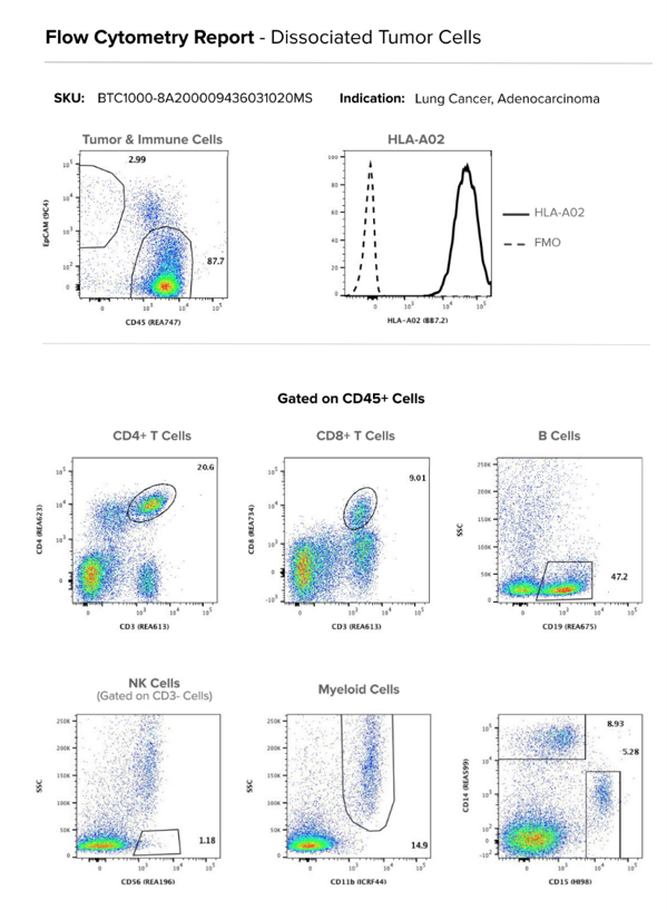
Figure 3. Example flow cytometry report for one lot of DTCs showing base flow cytometry profiles of cell populations, and high-resolution HLA-A02 status by flow cytometry
For isolation, CD45 (TIL) MicroBeads from Miltenyi Biotec enable efficient enrichment of CD45+ immune cells from dissociated tissue for downstream characterization and functional studies3. An important consideration is that TIL phenotype and function can be influenced by sample handling and processing workflows. Studies have shown that cryopreservation and repeated freeze-thaw cycles of human immune cell populations can affect immune cell recovery, surface marker expression, and functional readouts, potentially introducing variability into downstream analyses. When possible, downstream characterization should be performed immediately following TIL isolation to minimize additional sample handling and preserve biologically relevant immune characteristics.
For this reason, researchers often benefit from working with a partner that can provide well-characterized DTC biospecimens, TIL enrichment, and downstream characterization within a coordinated workflow. This integrated approach helps reduce pre-analytical variability and supports more consistent, reliable interpretation of immune profiling and functional assay results.
FFPE Tissue Provides Important Spatial Context
While DTCs provide access to viable TILs for isolation and functional studies, FFPE tissue provides complementary spatial context about where those TILs are located within the TME. Researchers can evaluate TIL localization within the tumor, including whether immune cells are infiltrating tumor regions or excluded from tumor regions and confined to surrounding stromal areas within the preserved tissue architecture. FFPE samples can also be used to characterize immune cell phenotypes, density, distribution, and activation states using immunohistochemistry and multiplex immunofluorescence. These spatial insights provide important context for understanding immune responses and identifying biomarkers associated with therapeutic response.
Moreover, digital pathology platforms, such as Discovery’s DiscoverAI, can further support scalable analysis of FFPE specimens by enabling quantitative evaluation of the tumor microenvironment, including spatial TIL characterization, and identifying biomarkers associated with therapeutic response.
Beyond Characterization: Using TILs to Model the Tumor Microenvironment
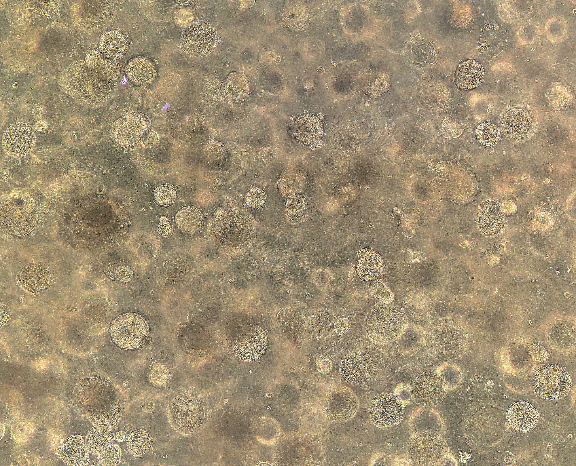
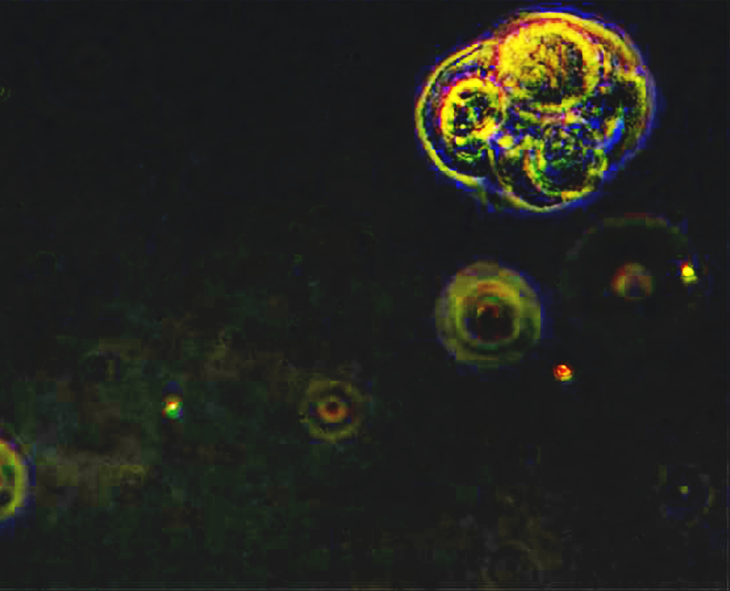
Beyond phenotypic and functional characterization, TILs isolated from DTCs can be incorporated into advanced in vitro models that more closely recapitulate the TME. For example, autologous TILs can be co-cultured with Discovery’s matched patient-derived 3D tumoroids to recreate key cellular interactions between tumor and immune cells while preserving patient-specific biology5. These models capture aspects of the cellular diversity and tumor-immune interactions of the parental tumor that are not represented in conventional monoculture systems.
As a result, TIL-tumoroid co-culture systems are emerging as physiologically relevant platforms for studying tumor-immune interactions, investigating mechanisms of immune evasion, and evaluating novel immuno-oncology therapies6. In one study, the addition of autologous TILs to breast cancer-derived tumoroids enhanced sensitivity to doxorubicin, demonstrating how these models can reveal treatment responses that may not be observed in cultures containing tumor cells alone7.
As immuno-oncology continues to evolve, the ability to study tumor-immune interactions in biologically relevant systems will be critical for developing the next generation of therapies. Combining well-characterized biospecimens, advanced immune profiling, and patient-derived models enables researchers to better understand immune responses, uncover mechanisms of response and resistance, and identify new opportunities to improve therapeutic outcomes.
Next in the Series: Cancer-Associated Fibroblasts
Stay tuned for the next blog in this series, where we will explore cancer-associated fibroblasts (CAFs), another important component of the TME. While TILs provide insights into the immune landscape within tumors, CAFs offer another perspective into the complex cellular interactions that influence tumor biology and disease progression.
References
- Kraja, F. P., Jurisic, V. B.,Hromić-Jahjefendić, A., Rossopoulou, N., Katsila, T., Mirjacic Martinovic, K., De Las Rivas, J., Diaconu, C. C., & Szöőr, Á. (2025). Tumor-infiltrating lymphocytes in cancer immunotherapy: from chemotactic recruitment to translational modeling. Frontiers in immunology, 16, 1601773. https://doi.org/10.3389/fimmu.2025.1601773
- Woodford, R., Lorigan, P., Oudit, D.,Abdulgawad, A., Thistlethwaite, F., & Lim, K. H. J. (2026). Tumour-infiltrating lymphocyte therapy in melanoma: ready for prime time?. British journal of cancer, 134(11), 1501–1509. https://doi.org/10.1038/s41416-026-03350-z
- Discovery Life Sciences.(2025). Dissociated Tumor and Tissue Cells User Guide: Best Practices For Handling DTCs And Preparation For Downstream Analysis. https://dls.com/resources/dtcs-user-guide/
- Serra, V., Fiorillo, E.,Cucca, F., & Orrù, V. (2022). Quantifying the Detrimental Effects of Multiple Freeze/Thaw Cycles on Primary Human Lymphocyte Survival and Function. International journal of molecular sciences, 24(1), 634. https://doi.org/10.3390/ijms24010634
- Su, Z., Li, H., Zhang, D., Shi, N., Song, C., Huang, Y., He, W., Yin, Z., & Li, L. (2026). Analysis of the tumor reactivity of autologous TILs and allogeneicγδT cells via tumor organoid-immune cell coculture. Journal of translational medicine, 24(1), 595. https://doi.org/10.1186/s12967-026-07706-0
- Wang, J., Tao, X., Zhu, J.,Dai, Z., Du, Y., Xie, Y., Chu, X., Fu, G., & Lei, Z. (2025). Tumor organoid-immune co-culture models: exploring a new perspective of tumor immunity. Cell Death Discovery, 11(1). https://doi.org/10.1038/s41420-025-02407-x
- Tokumaru, Y., Oshi, M., Katsuta, E., Matsuhashi, N., Futamura, M., Yoshida, K., &Takabe, K. (2020). Abstract 317: Organoid and TIL co-culture system models the human tumor immune microenvironment and drug sensitivity. Cancer Research, 80(16_Supplement), 317–317. https://doi.org/10.1158/1538-7445.am2020-317

Debbie King
Scientific Writer, Discovery Life Sciences